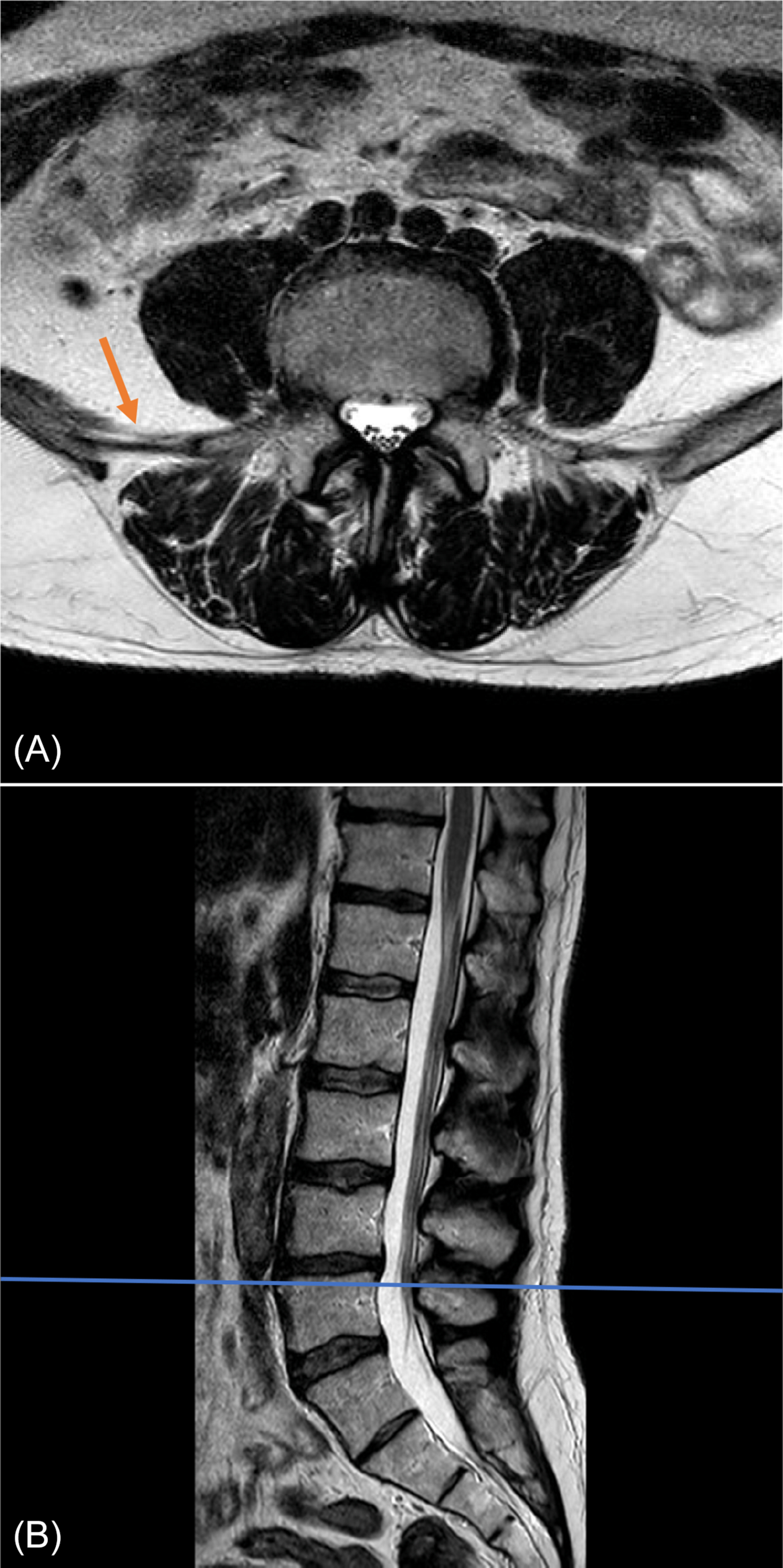
A lower back MRI (magnetic resonance imaging) is a diagnostic test that provides detailed images of the organs and tissues in the lower back region. The lower back, also known as the lumbar spine, is comprised of several structures, including the vertebrae, intervertebral discs, spinal cord, nerves, muscles, and blood vessels.
The MRI scan allows healthcare professionals to assess these structures and identify any abnormalities or conditions that may be causing lower back pain or discomfort. It provides a high-resolution view of the vertebrae, which are the bones that make up the spinal column. The MRI can detect fractures, tumors, or infections in these bones.
Furthermore, the intervertebral discs, which act as shock absorbers between the vertebrae, can be examined in detail. MRI images can identify degenerative changes, such as herniated discs or bulging discs, which may compress spinal nerves and cause pain. Spinal stenosis, a condition characterized by the narrowing of the spinal canal, can also be observed using an MRI.
In addition to the bone and disc structures, an MRI can show the spinal cord and nerve roots. These delicate structures can be evaluated for any signs of compression, inflammation, or tumors that may be contributing to lower back symptoms.
The muscles surrounding the spine can also be assessed through an MRI scan. Muscle tears, strains, or other abnormalities can be visualized, helping physicians determine the cause of lower back pain and tailor treatment options accordingly.
Lastly, the blood vessels in the lower back region can be imaged using contrast-enhanced MRI techniques. This can aid in the diagnosis of conditions such as vascular malformations or tumors.
In conclusion, a lower back MRI provides a comprehensive view of the organs and tissues in the lumbar spine region. It enables healthcare professionals to identify and diagnose various conditions that may be causing lower back pain or discomfort, leading to appropriate treatment plans for patients.
What will a lumbar MRI without contrast show?
With the help of an MRI lumbosacral spine, your doctor can see the vertebrae, spinal disks, spinal canal, and spinal cord. No contrast material is used in MRI without contrast.Nov 9, 2023

Do you go all the way in for a lumbar MRI?
Your whole body will typically be inside the machine, depending on what part of your back you are having imaged. If you are getting a lumbar MRI or a spinal MRI, you will be fully inside the MRI machine. Your spinal scan can take anywhere from 15-90 minutes.
Does a lumbar MRI show inflammation?
Some of the inconsistencies that a lumbar spine MRI may show include compression or inflammation of the spinal cord and adjacent nerves, degeneration of joints such as vertebral facet joints, disc herniation, infection of the discs, spinal cord and vertebrae, trauma to the tissues, and tumors.
What organs can be seen on a lumbar MRI?
Lumbar spine MR imaging may detect abnormalities of the kidneys, adrenal glands, liver, spleen, aorta and para-aortic regions, inferior vena cava, or the uterus and adnexal regions.